![]()
FAQ of the Month
NOW AVAILABLE
![]() FREE DOWNLOAD:Patient Radiation Handout
FREE DOWNLOAD:Patient Radiation Handout
About
Background
It is currently estimated that 62 million CT scans are obtained in the United States each year.1 While debated, a recent study suggests that radiation exposure from medical imaging may be responsible for 1-3% of cancers worldwide.2 In light of recent media coverage focusing on the increased risk of cancer from CT scans, patients and physicians have become more concerned about the increased use of medical imaging. Patients are asking their primary care providers and emergency room physicians for information about their risk. In 2004, Lee et al. concluded that “patients are not given information about the risks, benefits and radiation dose for a CT scan”.3 Additionally, this study found that both patients and physicians were “unable to provide accurate estimates of CT doses”.3
While the need for education in this area has clearly been established, there are no widely available resources that provide information to both patients and health care providers about the increased risk of cancer from medical imaging. X-RayRisk.com is an educational website that focuses on estimating this risk. One of the site’s main features is a web based calculator that allows users to track their imaging history and estimate their personal risk, while providing answers to frequently asked questions.
There are no published studies that prove the direct causality between medical imaging and increased cancer risk. Current data on radiation exposure and cancer risk is based on data from survivors of atomic bombs, nuclear accidents and the early use of x-rays. The assumed increased risk of cancer from low dose medical exposure (CT scans and x-rays) is based on individuals exposed to high doses (atomic bombs and nuclear accidents). The theory that the increased risk holds true at these lower doses is called the linear no threshold model, and is the currently adopted model for calculating radiation risk.
Great effort has been made throughout the medical community to ensure patient safety while providing quality diagnostic images. It is important to realize that in a properly performed individual exam, the potential health benefits almost always outweigh the potential risks of radiation exposure. Simply put, patients should not hesitate having a study if it is medically indicated. This site aims to provide accurate information for patients and health care providers to facilitate well-informed discussions about the increased risk of cancer from low dose radiation exposure.
The American College of Radiology (ACR) and the International Atomic Energy Agency (IAEA) both recommend hospitals monitor radiation exposure. It may be some time before all hospitals have the ability to track individual exposure. This site allows patients to log-in, create their own imaging record and generate an X-ray Risk Report with information about cancer risk.
Methods
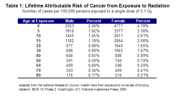
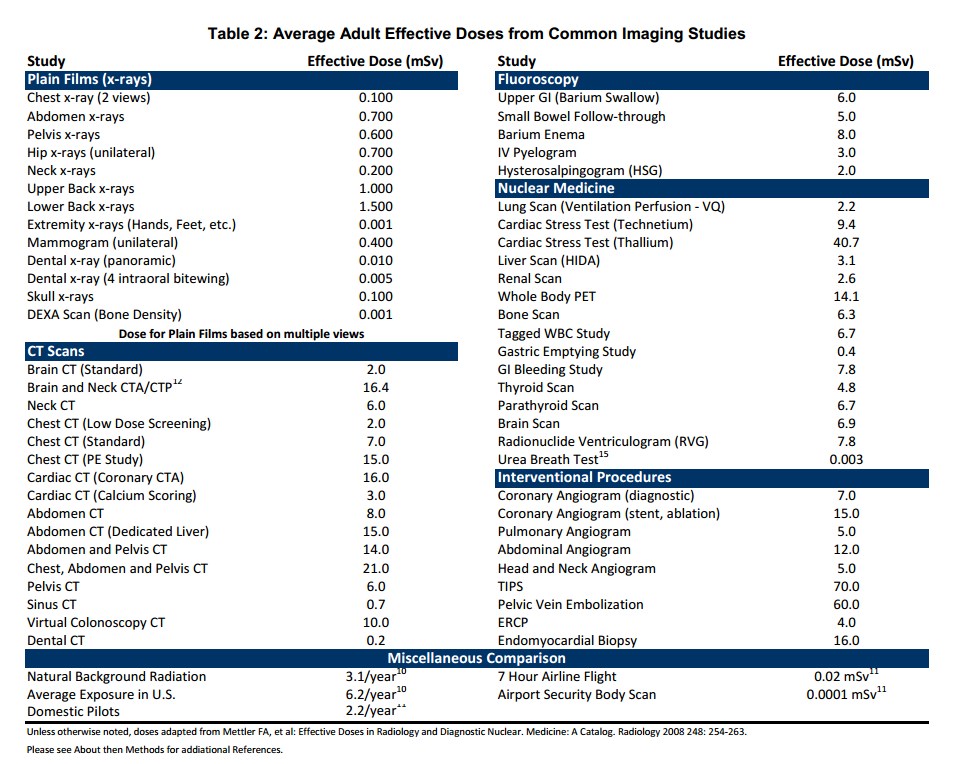
A committee of scientists and educators gathered by the National Research Council and organized by the National Academy of Sciences published their report on the Health Risks From Exposure to Low Levels of Ionizing Radiation in 2006. Table 1 was adapted from their Biological Effects of Ionizing Radiation (BEIR) VII Phase 2 Report. 4 The table estimates the number of additional cases of cancer attributable to a single dose of 0.1Gy (100 mSv) for different age groups. Data is based on the incidence of all cancer types. Data was plotted (Graph 1), exponential curves were drawn through the data points and formulas derived. Table 2 was adapted from Mettler et al and lists average adult doses for various medical imaging studies 5. The conversion factor used for the Brain and Neck CTA/CTP is 0.00345 12. The conversion factors used to Convert Your Dose from Dose Length Product (mGy · cm) to Effective Dose (mSv) were 0.0022 mSv/mGy · cm for Head CT, 0.0054 mSv/mGy · cm for Neck CT and 0.0180 mSv/mGy · cm for Body CT. 8 The Risk Calculator can also be used to convert Dose Length Product (DLP in mGy · cm) to Effective Dose (ED in mSv).
Click the images for an enlarged view
 |
 |
 |
Team
The content of this website has been reviewed by our medical team.
 |
Mike
Hanley, MD |
 |
Jay
Koonce, MD |
 |
Marques
Bradshaw, MD |
Resources
Image Wisely - Radiation Safety in Adult Medical Imaging (www.imagewisely.org)
Image Gently - The Alliance for Radiation Safety in Pediatric Imaging (www.imagegently.org)
Nuffield Health - Educational Radiation Video (https://www.youtube.com/watch?v=3Ik3Ej27Yp0)
Health Physics Society - Specialists in Radiation Protection (www.HPS.org)
RadiologyInfo.org - Radiology information resource for patients (www.radiologyinfo.org)
References
1. Brenner DJ, Hall EJ. Computed Tomography – An Increasing Source of Radiation Exposure. NEJM 357: 2277-84, 2007. (http://www.ncbi.nlm.nih.gov/pubmed/18046031)
2. Berrington de Gonzalez A, Darby S. Risk of cancer from diagnostic x-rays: estimates for the UK and 14 other countries. Lancet 2004; 363:345-51. (http://www.ncbi.nlm.nih.gov/pubmed/?term=15070562)
3. Lee CI, Haims AH, Monico EP, et al. Diagnostic CT Scans: Assessment of Patient, Physician, and Radiologist Awareness of Radiation Dose and Possible Risks. Radiology 231 (2): 393-398. (2004). (http://www.ncbi.nlm.nih.gov/pubmed/15031431)
4. National Research Council. Health risks from exposure to low levels of ionizing radiation. BEIR VII Phase 2. Washington, DC: National Academies Press; 2006.
5. Mettler FA, Huda W, Yoshizumi TT, Mahesh M: "Effective Doses in Radiology and Diagnostic Nuclear Medicine: A Catalog." Radiology 2008 248: 254-263. (http://www.ncbi.nlm.nih.gov/pubmed/18566177)
6. American Cancer Society: Cancer Facts and Figures 2008. 2008CAFFfinalsecured.pdf Cancer Incidence)
7. Office of Communications and Public Liaison and the Radiation Safety Branch of the Office of the Director, National Institutes of Health. Fact Sheet: What We Know About Radiation. Available at http://www.nih.gov/health/chip/od/radiation/. Accessed December 16, 2008
8. Huda W, Ogden KM, Khorasani MR: Converting Dose-Length Product to Effective Dose at CT. Radiology 248:995-1003, 2008. (http://www.ncbi.nlm.nih.gov/pubmed/18710988)
9. Brody AS, Frush DP, Huda W, et al: Radiation Risk to Children From Computed Tomography. Pediatrics, 120 (3): 677-682, 2007 (http://www.ncbi.nlm.nih.gov/pubmed/17766543)
10. NCRP Report No. 160, Ionizing Radiation Exposure of the Population of the United States. Available at www.ncrponline.org. Accessed April 2009.
11. Health Physics Society: Radiation Exposure During Commercial Airline Flights. Available at http://www.hps.org/publicinformation/ate/faqs/commercialflights.html. Accessed September 2009.
12. Mnyusiwalla A, Aviv RI, Symons SP: Radiation dose from multidector row CT imaging for acute stroke. Neuroradiology 51:635-640, 2009. (http://www.ncbi.nlm.nih.gov/pubmed/19506845)
13. McCollough CH, Guimaraes L, Fletcher JG: In Defense of Body CT. AJR 193:28-39, 2009. (http://www.ncbi.nlm.nih.gov/pubmed/19542392)
14. ACR Statement on Airport Full-body Scanners and Radiation, January 2010. (http://acr.org/MainMenuCategories/media_room/FeaturedCategories/PressReleases/StatementonAirportFullbodyScanners.aspx)
15. Balon HR, Roff E, Freitas JE, et al: Society of Nuclear Medicine Procedure Guideline for C-14 Urea Breath Test. Version 3.0, June 23, 2001 (http://interactive.snm.org/docs/pg_ch07_0403.pdf)
16. Calicchia A, Chiacchiararelli L, DeFelice C, et al: Assessment of radiation dose to patients in hysterosalpingography. Radiol Med 95(1-2): 93-7, 1998.
17. Platts D, Brown M, Javorsky G, et al: Comparison of fluoroscopic versus real-time three-dimensional transthoracic echocardiographic guidance of endomyocardial biopsies. Eur J of Echocardiography 11, 637-643, 2010. (http://www.ncbi.nlm.nih.gov/pubmed/20335406)
18. Hendrick ER: Radiation Doses and Cancer Risks from Breast Imaging. Radiology, 257 (1), 246- 253, 2010." (http://www.ncbi.nlm.nih.gov/pubmed/20736332)
19. Heusch P, Kropil P, Buchbender C, et al: Radiation exposure of the radiologist’s eye lens during CT-guided interventions. Acta Radiologica 2014, Vol 55(1) 86-90. (http://www.ncbi.nlm.nih.gov/pubmed/23884839)
20. Richards PJ, George J, Metelko M, et al: Spine computed tomography doses and cancer induction: Spine 35(4), 430-433 2010. (http://www.ncbi.nlm.nih.gov/pubmed/20081559)
Citations
In addition of educating patients and providers about the risks of radiation, we have designed our online calculator as a research tool for those researching radiation induced cancers. We are proud that since 2009 the XrayRisk.com website has been cited in 112 publications. Here are some select English language publications:
2021
Rose, L., Williams, R., Al-Ahmed, S., Fenner, C., Fragkakis, A., Lupu, C., ... & Lui, D. F. (2021, September). EOS low-dose radiation imaging for spinal deformity: The new gold standard. In Orthopaedic Proceedings (Vol. 103, No. SUPP_11, pp. 12-12). The British Editorial Society of Bone & Joint Surgery.
https://online.boneandjoint.org.uk/doi/abs/10.1302/1358-992X.2021.11.012
Pasternak, J. J. (2021). Radiation Exposure and the Neuroanesthesiologist. Journal of Neuroanaesthesiology and Critical Care, 8(01), 001-002.
https://www.thieme-connect.com/products/ejournals/html/10.1055/s-0041-1725229
Ford, B., Dore, M., & Moullet, P. (2021). Diagnostic imaging: appropriate and safe use. American Family Physician, 103(1), 42-50.
https://www.aafp.org/afp/2021/0101/p42
Morrison, N., Bryden, S., & Costa, A. F. (2021). Split vs. Single Bolus CT Urography: Comparison of Scan Time, Image Quality and Radiation Dose. Tomography, 7(2), 210-218.
https://www.mdpi.com/2379-139X/7/2/19
Keller, G., Afat, S., Ahrend, M. D., & Springer, F. (2021). Diagnostic accuracy of ultra-low-dose CT for torsion measurement of the lower limb. European Radiology, 31(6), 3574-3581.
https://link.springer.com/article/10.1007/s00330-020-07528-8
Weinstein, O., Yitshak Sade, M., Shelef, I., Novack, V., Abu Tailakh, M., & Levy, J. (2021). The association between exposure to radiation and the incidence of cataract. International Ophthalmology, 41(1), 237-242.
https://link.springer.com/article/10.1007/s10792-020-01572-5
Caffrey, E. (2021). Radiation and the skeptical public: tips and tools for communicating effectively. Health Physics, 120(6), 693-698.
Loughenbury, P. R., Gentles, S. L., Murphy, E. J., Tomlinson, J. E., Borse, V. H., Dunsmuir, R. A., ... & Khan, A. L. (2021). Estimated cumulative X-ray exposure and additional cancer risk during the evaluation and treatment of scoliosis in children and young people requiring surgery. Spine Deformity, 9(4), 949-954.
https://link.springer.com/article/10.1007/s43390-021-00314-6
Keller, G., Götz, S., Kraus, M. S., Grünwald, L., Springer, F., & Afat, S. (2021). Radiation Dose Reduction in CT Torsion Measurement of the Lower Limb: Introduction of a New Ultra-Low Dose Protocol. Diagnostics, 11(7), 1209.
2020
Atiyyah, T. A. E. R., Nasr, M. S. N., Ahmed, T. S., & Mostafa, M. M. S. A. (2020). Cumulative Radiation Exposure from Diagnostic Imaging in Zagazig University Pediatric Intensive Care and Chest Units. The Egyptian Journal of Hospital Medicine, 81(2), 1520-1524.
https://journals.ekb.eg/article_115566.html
Abuzaid, M. M., Elshami, W., Bukamal, B., Alawnih, L., Sabeel, W., & Noorajan, Z. (2020, January). Correlation between radiation-induced cancer and the accumulative dose for the patient underwent multiple brain CT scan. European Congress of Radiology-ECR 2020.
Spiliopoulos, S., Reppas, L., Zompola, C., Palaiodimou, L., Papadopoulou, M., Filippiadis, D., ... & Brountzos, E. (2020). Computed‐tomography‐guided transforaminal intrathecal nusinersen injection in adults with spinal muscular atrophy type 2 and severe spinal deformity. Feasibility, safety and radiation exposure considerations. European Journal of Neurology, 27(7), 1343-1349.
https://onlinelibrary.wiley.com/doi/abs/10.1111/ene.14245
Abend, M., Stricklin, D., Flaig, N., Badie, C., Drouet, M., Foster, C., ... & Port, M. (2020). Bringing Radiation Exposures and Associated Health Risks into Perspective—Development of an App. Health Physics, 119(1), 59-63.
Borgbjerg, J., Bylling, T., Andersen, G., Thygesen, J., Mikkelsen, A., & Nielsen, T. K. (2020). CT-guided cryoablation of renal cancer: radiation burden and the associated risk of secondary cancer from procedural-and follow-up imaging. Abdominal Radiology, 45(11), 3581-3588.
https://link.springer.com/article/10.1007/s00261-020-02527-1
Luís, A., Navarro-Ramirez, R., Kirnaz, S., Nakhla, J., & Härtl, R. (2020). Navigated Spinal Fusion 31. Minimally Invasive Spine Surgery: Surgical Techniques and Disease Management, 355.
2019
Lewis, S., Young, B., Thurley, P., Shaw, D., Cranwell, J., Skelly, R., ... & Fogarty, A. (2019). Evaluation of a nudge intervention providing simple feedback to clinicians of the consequence of radiation exposure on demand for computed tomography: a controlled study. Clinical Medicine, 19(4), 290.
https://www.ncbi.nlm.nih.gov/pmc/articles/PMC6752234/
Banerjee, P., & Thomas, M. (2019). CT scans to exclude spine fractures in children after negative radiographs may lead to increase in future cancer risk. European Journal of Orthopaedic Surgery & Traumatology, 29(5), 983-988.
https://link.springer.com/article/10.1007/s00590-019-02396-5
Liberman, D. B., & McCarthy, T. J. (2019). The cost of callbacks: return visits for diagnostic imaging discrepancies in a pediatric emergency department. Emergency Radiology, 26(4), 381-389.
https://link.springer.com/article/10.1007/s10140-019-01681-4
Cordiner, D., Al-Ani, M., Jia, X., Marchick, M., Allen, B., & Winchester, D. (2019). Estimates of radiation exposure and subsequent risk of malignancy due to cardiac imaging in the emergency department for evaluation of chest pain: a cohort study. Coronary artery disease, 30(8), 626.
https://www.ncbi.nlm.nih.gov/pmc/articles/PMC6832827/
McKenney, S., Gingold, E., & Zaidi, H. (2019). Gonadal shielding should be discontinued for most diagnostic imaging exams. Medical Physics, 46(3), 1111-1114.
https://archive-ouverte.unige.ch/unige:115014
Skelly, R., Sturrock, N., Norwood, M., & Thurley, P. (2019). Evaluation of the impact of a brief educational message on clinicians' awareness of risks of ionising-radiation exposure in imaging investigations: a pilot pre-post intervention study.
Gofrit, O., & Orevi, M. (2019). Post-operative surveillance in kidney cancer. Annals of Translational Medicine, 7(Suppl 3).
https://www.ncbi.nlm.nih.gov/pmc/articles/PMC6685866/
Wurster, C. D., Winter, B., Wollinsky, K., Ludolph, A. C., Uzelac, Z., Witzel, S., ... & Kocak, T. (2019). Intrathecal administration of nusinersen in adolescent and adult SMA type 2 and 3 patients. Journal of neurology, 266(1), 183-194.
https://link.springer.com/article/10.1007/s00415-018-9124-0
Adam, A. H. Y. (2019). Assessment of Radiation Dose to Head, Chest and Abdomen of Adult Patients Underwent Computed Tomography Examination-Khartoum State-Sudan (Doctoral dissertation, Sudan University of Science and Technology).
http://repository.sustech.edu/handle/123456789/24474
Young, B., Cranwell, J., Fogarty, A. W., Skelly, R., Sturrock, N., Norwood, M., ... & Thurley, P. (2019). Evaluation of the impact of a brief educational message on clinicians’ awareness of risks of ionising-radiation exposure in imaging investigations: a pilot pre-post intervention study. BMC Health Services Research, 19(1), 1-7.
https://bmchealthservres.biomedcentral.com/articles/10.1186/s12913-019-4712-y
Lin, E., & Schueler, B. (2019). Radiologic issues and radiation safety during ERCP. In ERCP (pp. 14-29). Elsevier.
https://www.sciencedirect.com/science/article/pii/B9780323481090000031
Luís, A., Navarro-Ramirez, R., Kirnaz, S., Nakhla, J., & Härtl, R. (2019). Navigated spinal fusion. In Minimally Invasive Spine Surgery (pp. 355-374). Springer, Cham.
https://link.springer.com/chapter/10.1007/978-3-030-19007-1_31
2018
Weiss, E. S. (2018). Estimating cancer risk from radiation. Canadian Family Physician, 64(1), 8.
https://www.ncbi.nlm.nih.gov/pmc/articles/PMC5962978/
Wray, C. M., & Cho, H. J. (2018). Annals for Hospitalists Inpatient Notes-Medical Uncertainty as a Driver of Resource Use—Examining the “Gray Zones” of Clinical Care. Annals of Internal Medicine, 168(12), HO2-HO3.
https://www.acpjournals.org/doi/full/10.7326/M18-1320
Maclean, D., Maher, B., Harris, M., Dyer, J., Modi, S., Hacking, N., & Bryant, T. (2018). Planning prostate artery embolisation: is it essential to perform a pre-procedural CTA?. CardioVascular and Interventional Radiology, 41(4), 628-632.
https://link.springer.com/article/10.1007/s00270-017-1842-7
Corson-Knowles, D., & Russell, F. M. (2018). Clinical ultrasound is safe and highly specific for acute appendicitis in moderate to high pre-test probability patients. Western Journal of Emergency Medicine, 19(3), 460.
https://www.ncbi.nlm.nih.gov/pmc/articles/PMC5942008/
Viceconti, M., Qasim, M., Bhattacharya, P., & Li, X. (2018). Are CT-based finite element model predictions of femoral bone strengthening clinically useful?. Current osteoporosis reports, 16(3), 216-223.
https://link.springer.com/article/10.1007/s11914-018-0438-8
Paranathala, M. P., Ferguson, L., Bowers, R., & Mukerji, N. (2018). Percutaneous retrogasserian glycerol rhizotomy for trigeminal neuralgia: an alternative technique. British Journal of Neurosurgery, 32(6), 657-660.
https://www.tandfonline.com/doi/abs/10.1080/02688697.2018.1504882
Rubai, S. S., Rahman, M. S., Purohit, S., Patwary, M. K. A., Moinul, A. K. M., Meaze, H., & Mamun, A. A. (2018). Measurements of Entrance Surface Dose and Effective Dose of Patients in Diagnostic Radiography. Biomedical Journal, 1, 5.
https://www.academia.edu/download/84709574/BJSTR.MS.ID.002186.pdf
Gofrit, O. N., Rabinovich, I., Yutkin, V., Pode, D., Duvdevani, M., Landau, E. H., ... & Goldberg, S. N. (2018, November). Abbreviated CT protocol for postoperative surveillance of renal cancer. In Urologic Oncology: Seminars and Original Investigations (Vol. 36, No. 11, pp. 498-e9). Elsevier.
https://www.sciencedirect.com/science/article/pii/S1078143918303028
Chang, C. Y., Simeone, F. J., DeLorenzo, M. C., Palmer, W. E., Bredella, M. A., & Huang, A. J. (2018). Dose reduction for fluoroscopically guided injections: phantom simulation and patient procedures. Skeletal Radiology, 47(2), 223-231.
https://link.springer.com/article/10.1007/s00256-017-2788-1
Hsiao, V., Santillanes, G., Malek, D., & Claudius, I. (2018). Review of interventions and radiation exposure from chest computed tomography in children with blunt trauma. The Journal of Pediatrics, 198, 220-225.
https://www.sciencedirect.com/science/article/pii/S002234761830338X
Levsky, J. M., Haramati, L. B., Spevack, D. M., Menegus, M. A., Chen, T., Mizrachi, S., ... & Garcia, M. J. (2018). Coronary computed tomography angiography versus stress echocardiography in acute chest pain: a randomized controlled trial. JACC: Cardiovascular Imaging, 11(9), 1288-1297.
https://www.jacc.org/doi/abs/10.1016/j.jcmg.2018.03.024
Resnik, D. B. (2018). Risks. In The Ethics of Research with Human Subjects (pp. 165-191). Springer, Cham.
https://link.springer.com/chapter/10.1007/978-3-319-68756-8_7
2017
Nyheim, T., Staxrud, L. E., Jørgensen, J. J., Jensen, K., Olerud, H. M., & Sandbæk, G. (2017). Radiation exposure in patients treated with endovascular aneurysm repair: what is the risk of cancer, and can we justify treating younger patients?. Acta Radiologica, 58(3), 323-330.
https://journals.sagepub.com/doi/abs/10.1177/0284185116651002
Leech, C. (2017). Whole body computed tomography for trauma: friend or foe?. Emergency Medicine Journal, 34(10), 635-636.
https://emj.bmj.com/content/34/10/635.abstract
Maclean, D., Maher, B., Modi, S., Harris, M., Dyer, J., Somani, B., ... & Bryant, T. (2017). Prostate artery embolization: a new, minimally invasive treatment for lower urinary tract symptoms secondary to prostate enlargement. Therapeutic Advances in Urology, 9(8), 209-216.
https://journals.sagepub.com/doi/abs/10.1177/1756287217717889
Lukoff, J., & Olmos, J. (2017). Minimizing medical radiation exposure by incorporating a new radiation “vital sign” into the electronic medical record: quality of care and patient safety. The Permanente Journal, 21.
https://www.ncbi.nlm.nih.gov/pmc/articles/PMC5638638/
Tsiouris, A. J., Qian, J., & Kummer, B. R. (2017). Advanced Imaging in Acute Ischemic Stroke. Emergency Medicine Reports, 38(24).
2016
Mendelson, R. M., & Montgomery, B. D. (2016). Towards appropriate imaging: tips for practice. Australian Family Physician, 45(6), 391-395.
https://search.informit.org/doi/abs/10.3316/INFORMIT.138702282271638
Kinsara, A., El-Gizawy, A. S., Banoqitah, E., & Ma, X. (2016). Review of Leakage from a Linear Accelerator and Its Side Effects on Cancer Patients J Nucl Med Radiat Ther 7: 288. doi: 10.4172/2155-9619.1000288 Page 2 of 6 Volume 7• Issue 3• 1000288 J Nucl Med Radiat Ther ISSN: 2155-9619
Wanek, J., & Rühli, F. J. (2016). Risk to fragmented DNA in dry, wet, and frozen states from computed tomography: a comparative theoretical study. Radiation and environmental biophysics, 55(2), 229-241.
https://link.springer.com/article/10.1007/s00411-016-0637-6
Dalman, R. L., & Mell, M. (2016). Management of asymptomatic abdominal aortic aneurysm. Official reprint from UpToDate.[Google Scholar].
https://www.medilib.ir/uptodate/show/15188
St Jacques, M. A. (2016). Their swords, our plowshares:" Peaceful" nuclear weapons, propaganda, and Cold War memory expressed in film: 1959-1989.
https://commons.lib.jmu.edu/cgi/viewcontent.cgi?article=1105&context=master201019
2015
Eylert, G., Deutinger, M., Stemberger, A., Huber, W., & Gösseringer, N. (2015). Evaluation of the perforator CT-angiography with a cancer risk assessment in DIEP flap breast reconstruction. Journal of Plastic, Reconstructive & Aesthetic Surgery, 68(4), e80-e82.
https://www.jprasurg.com/article/S1748-6815(14)00736-0/abstract
Lam, D. L., Larson, D. B., Eisenberg, J. D., Forman, H. P., & Lee, C. I. (2015). Communicating potential radiation-induced cancer risks from medical imaging directly to patients. American Journal of Roentgenology, 205(5), 962-970.
https://www.ajronline.org/doi/abs/10.2214/AJR.15.15057
Semghouli, S., Amaoui, B., & Maamri, A. (2015). Estimated radiation exposure from medical imaging for patients of radiology service of Al Faraby Hospital, Oujda Morocco. International Journal of Cancer Therapy and Oncology, 3(3), 3325.
Logsdon, R., & Gleason, R. (2015). Advanced nurse practitioner educational needs for safe and efficient radiological imaging. Advanced Emergency Nursing Journal, 37(3), 233-241.
Hikino, K., & Yamamoto, L. G. (2015). The benefit of neck computed tomography compared with its harm (risk of cancer). Journal of Trauma and Acute Care Surgery, 78(1), 126-131.
Levsky, J. M., Spevack, D. M., Travin, M. I., Menegus, M. A., Huang, P. W., Clark, E. T., ... & Haramati, L. B. (2015). Coronary computed tomography angiography versus radionuclide myocardial perfusion imaging in patients with chest pain admitted to telemetry: a randomized trial. Annals of internal medicine, 163(3), 174-183.
https://www.acpjournals.org/doi/abs/10.7326/m14-2948
2014
Saef, S. H., Hanley, M., Zhao, W., Walker, B. J., & Carr, C. M. (2014). 157 Potential to Reduce Lifetime Attributable Risk of Cancer through Avoidance of Radiographic Imaging in the Emergency Department by using a Health Information Exchange: An Observational Study Based on Emergency Department Clinician Perceptions. Annals of Emergency Medicine, 64(4), S57.
https://www.annemergmed.com/article/S0196-0644(14)00790-2/abstract
Wang, Y. X. J., Lo, G. G., Yuan, J., Larson, P. E., & Zhang, X. (2014). Magnetic resonance imaging for lung cancer screen. Journal of thoracic disease, 6(9), 1340.
https://www.ncbi.nlm.nih.gov/pmc/articles/PMC4178109/
Kashcheev, V. V., Pryakhin, E. A., Menyaylo, A. N., Chekin, S. Y., & Ivanov, V. K. (2014). Comparing risk estimates following diagnostic CT radiation exposures employing different methodological approaches. Health Physics, 106(6), 806-811.
Wang, Y. X. J., Gong, J. S., Suzuki, K., & Morcos, S. K. (2014). Evidence based imaging strategies for solitary pulmonary nodule. Journal of thoracic disease, 6(7), 872.
https://www.ncbi.nlm.nih.gov/pmc/articles/PMC4120179/
Bermea–Mendoza, J. H., Franco–Herbert, A., Onofre–Castillo, J. J., Valero–Castillo, R., & Alba–Quintanilla, D. (2014). Dose of radiation given to patient in intensive care. In Anales de Radiología, México (Vol. 13, No. 1, pp. 45-52).
https://www.medigraphic.com/cgi-bin/new/resumenI.cgi?IDARTICULO=48522
Ho, I. K., Cash, B. D., Cohen, H., Hanauer, S. B., Inkster, M., Johnson, D. A., ... & Quigley, E. M. (2014). Radiation exposure in gastroenterology: improving patient and staff protection. Official journal of the American College of Gastroenterology| ACG, 109(8), 1180-1194.
2013
Skinner, S. (2013). Radiation safety. Australian family physician, 42(6), 387-389.
https://search.informit.org/doi/abs/10.3316/informit.359954294780783
Willoughby, H., Ahmed, H., Jenkinson, R., & Edwards, A. (2013). GP speciality trainees’ knowledge, attitude and practice regarding risks associated with common radiological investigations. Education for Primary Care, 24(5), 355-362.
https://www.tandfonline.com/doi/pdf/10.1080/14739879.2013.11494200
Ramanathan, S., & Ryan, J. Radiation awareness among radiology residents, fellows, staff and technologists.
Ivanov, V. K., Kashcheev, V. V., Chekin, S. Y., Menyaylo, A. N., Pryakhin, E. A., Tsyb, A. F., & Mettler, F. A. (2013). Estimation of risk from medical radiation exposure based on effective and organ dose: how much difference is there?. Radiation protection dosimetry, 155(3), 317-328.
https://academic.oup.com/rpd/article-abstract/155/3/317/1612109
Vazquez, J. L., Pombar, M. A., Pumar, J. M., & Del Campo, V. M. (2013). Optimised low-dose multidetector CT protocol for children with cranial deformity. European radiology, 23(8), 2279-2287.
https://link.springer.com/article/10.1007/s00330-013-2806-1
Latifi, A. (2013). Exploring new protocols in emergency radiology, and their impact on radiation and diagnosis. Karolinska Institutet (Sweden).
Adhikari, S. R. (2013). Plexar Imaging: A Startup Determined to Solve the CT Dose Variability Problem (Doctoral dissertation, Case Western Reserve University).
https://rave.ohiolink.edu/etdc/view?acc_num=case1374236161
2012
Weltz, A. (2012). QUICK RADIATION DOSE RESOURCE FOR PATIENTS. Health Physics, 103(2).
Carpeggiani, C., Paterni, M., Caramella, D., Vano, E., Semelka, R. C., & Picano, E. (2012). A novel tool for user-friendly estimation of natural, diagnostic and professional radiation risk: Radio-Risk software. European journal of radiology, 81(11), 3563-3567.
https://www.sciencedirect.com/science/article/pii/S0720048X11006061
Johnson, J. O., & Robins, J. M. (2012). CT imaging: radiation risk reduction—real-life experience in a metropolitan outpatient imaging network. Journal of the American College of Radiology, 9(11), 808-813.
https://www.sciencedirect.com/science/article/pii/S1546144012003766
De Gonzalez, A. B., Apostoaei, A. I., Veiga, L. H., Rajaraman, P., Thomas, B. A., Hoffman, F. O., ... & Land, C. (2012). RadRAT: a radiation risk assessment tool for lifetime cancer risk projection. Journal of Radiological Protection, 32(3), 205.
https://iopscience.iop.org/article/10.1088/0952-4746/32/3/205/meta
Gerstenmaier, J. F. (2012). Public health implications of medical diagnostic radiation exposure (Doctoral dissertation).
https://uir.unisa.ac.za/handle/10500/6085
2011
Davies, H. E., Wathen, C. G., & Gleeson, F. V. (2011). The risks of radiation exposure related to diagnostic imaging and how to minimise them. Bmj, 342.
https://www.bmj.com/content/342/bmj.d947.extract
Stamm, G. (2011). Software for Calculating Dose and Risk. In Radiation Dose from Multidetector CT (pp. 563-568). Springer, Berlin, Heidelberg.
https://link.springer.com/chapter/10.1007/174_2011_434
2010
Alessio, A. M., & Phillips, G. S. (2010). A pediatric CT dose and risk estimator. Pediatric radiology, 40(11), 1816-1821.
https://link.springer.com/article/10.1007/s00247-010-1761-0
White, H. A., & Macdonald, S. (2010). Estimating risk associated with radiation exposure during follow-up after endovascular aortic repair (EVAR). Journal of cardiovascular surgery, 51(1), 95.
| Refer this site | Updated on 2024-03-28 | Terms & Conditions | Privacy Policy |
Together, dedicated to improving the understanding of radiation risks from medical imaging. Calculate your dose and estimate cancer risk from studies including CT scans, x-rays, nuclear scans and interventional procedures. |