FAQ’s
Where does radiation come from?
How do x-rays increase your risk for cancer?
Why isn’t there a study directly linking medical imaging and cancer?
Isn’t radiation used to treat cancer?
If I have cancer, can radiation from medical imaging make it worse?
Why is the average risk of developing cancer so high?
Why does age and gender matter when calculating cancer risk?
If I am pregnant, what is the risk to the fetus?
If x-rays increase my risk of cancer, why should I get a Mammogram to screen for cancer?
Does MRI or Ultrasound cause cancer?
How does the risk from medical radiation compare to background radiation exposure?
How does airline travel expose me to radiation?
How do I protect myself from radiation exposure?
Does the Government regulate radiation exposure?
How much radiation do airport security whole body scanners use?
Do mammograms cause an increase in thyroid cancer?
What is radiation?
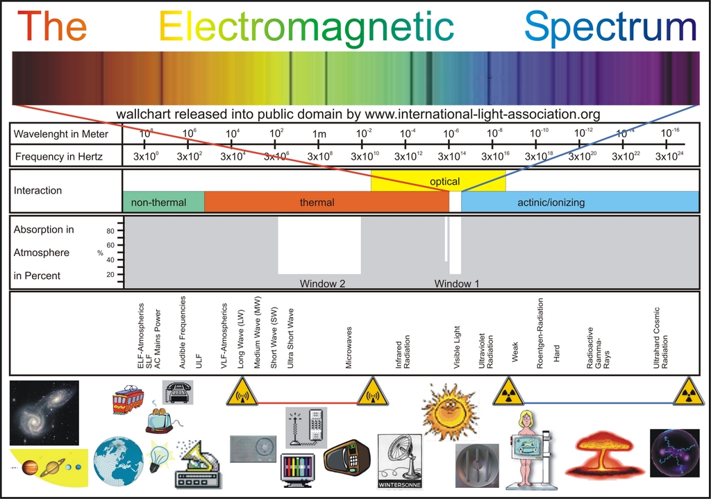 Radiation can simply be described as energy moving through space. It can
take many forms, including visible light, x-rays,
gamma-rays, microwaves and radio waves. This site specifically addresses
high energy or ionizing radiation, which includes x-rays.
Ionizing radiation has many uses, including sterilization of food and medical
equipment, creation of medical images, and is even used in the treatment
Radiation can simply be described as energy moving through space. It can
take many forms, including visible light, x-rays,
gamma-rays, microwaves and radio waves. This site specifically addresses
high energy or ionizing radiation, which includes x-rays.
Ionizing radiation has many uses, including sterilization of food and medical
equipment, creation of medical images, and is even used in the treatment
Where does radiation come from?
Radiation is all around us. Currently, two main sources of ionizing radiation are from natural background radiation and medical exposure (CT scans and x-rays). Natural background radiation comes from the Sun (cosmic radiation), the Earth (mostly Radon gas) and from naturally radioactive substances in our body. Natural background radiation exposure accounts for an average of 3.1 mSv/yr with variations depending on where you live.
What are x-rays?
X-rays are a type of radiation that are created using large amounts of electricity. X-rays are used in medical imaging much like a camera uses visible light to create an image. X-rays pass through the body and create an image on film based on how many x-rays get absorbed and how many pass through. These films are commonly referred to as “x-rays,” but x-rays are actually the type of radiation that is used to produce the image. Studies that use x-rays include plain films, fluoroscopy and computed tomography (CT scans).
How do x-rays increase your risk for cancer?
When x-rays, or any ionizing radiation, pass through the body they cause electrons to be ejected from atoms, leaving behind positive ions. These positive ions, or free radicals, can cause damage to DNA. DNA can also be damaged directly by radiation. If DNA is damaged, there are three possible outcomes:
- The cell dies (only occurs with very high doses).
- The cell repairs itself perfectly (most common result).
- The cell repairs itself with mistakes (rare).
The inaccurate repair of DNA is rare, but can cause a cell to act wildly or grow into a cancer. Oftentimes it takes decades for cancer to be detected following radiation exposure.
Why isn’t there a study directly linking medical imaging and cancer?
There are no studies that directly link cancer to the low dose radiation used in current medical imaging. To scientifically prove a connection would require nearly one million patients followed closely over decades to detect the small increased risk with any confidence.
Isn’t radiation used to treat cancer?
Radiation is very successful at treating some cancers. An entire field of medicine (Radiation Oncology) is devoted to this practice. Radiation treatment doses are much higher than doses used for medical imaging. High dose radiation causes cell death, specifically the cells which are growing the fastest including cancer cells, hair cells and gastrointestinal tract lining.
If I have cancer, can radiation from medical imaging make it worse?
No. Low dose radiation from medical imaging does not affect known cancer. In fact, high dose radiation is used to treat cancer. Low dose exposure increases the risk of developing new cancer decades after exposure, which is the focus of this site. The information gained from imaging patients with cancer most likely outweighs the small risk of cancer induction many years in the future.
Why is the average risk of developing cancer so high?
Regardless of radiation exposure, the average overall lifetime risk of developing an invasive cancer is 37.5% for women and 44.9% for men.6 These statistics are averages and do not predict what is going to happen to you. They do not take into consideration individual risk factors including lifestyle (smoking, diet, exercise, etc.), family history (genetics) or radiation exposure. The majority of cancers occur later in life and the average lifetime risk of dying from cancer is 25%.6
Why does age and gender matter when calculating cancer risk?
Pediatric patients are at the greatest risk of developing cancer from radiation exposure. There are two theories why. First, rapidly dividing or growing cells are at greater risk of damage from ionizing radiation. Second, children have a long life ahead of them, therefore the chance of detecting a slow growing cancer is higher when compared to someone exposed later in life. With this in mind, most institutions make adjustments in the way they take images of pediatric patients, using lower doses or shielding sensitive organs.
In general, women are at slightly higher risk of developing cancer when compared to men exposed to the same dose of radiation. This is based on high dose exposure data from survivors of atomic bombs, nuclear accidents and the early use of x-rays. Men and women also have different average risks for developing cancer.
If I am pregnant, what is the risk to the fetus?
Much like the risk estimates in adults, fetal risk estimates are not proven with any certainty, but are taken very seriously. We know that children are more sensitive than adults, so we assume the fetus is at even higher risk. If there is any chance you may be pregnant, you must inform your doctor as well as the staff that is performing your study. There are precautions that can be taken to limit risk or there may be alternative imaging modalities, such as MRI and Ultrasound. The estimates provided by this website are not designed to estimate fetal risk.
If x-rays increase my risk of cancer, why should I get a Mammogram to screen for cancer?
Carefully evaluating the risks and benefits of screening studies (like mammograms) is an important part of medicine. Screening mammograms have been proven to decrease mortality from breast cancer by approximately 30%. Simply put, early detection of breast cancer using mammograms saves lives. Therefore, the American Cancer Society recommends an annual screening mammogram for women over age 40. The risk of getting cancer from the mammogram itself is negligible. Women at high risk for breast cancer should discuss imaging options with their doctor.
Does MRI or Ultrasound cause cancer?
MRI uses strong magnetic fields and radio waves to obtain images, which have not been associated with an increased risk of cancer. Ultrasound uses sound waves to produce images. There is no exposure to ionizing radiation with MRI or Ultrasound.
How does the risk from medical radiation compare to background radiation exposure?
Natural background radiation exposure accounts for an average of 3.1 mSv/yr with variations depending on where you live. In the US, the average person is exposed to an additional 3.0 mSv/yr from medical sources (predominantly CT scans). Of course, some people receive no radiation and others much, much more. The average US total radiation exposure (all sources) is 6.2 mSv/yr which is an increase from 20 years ago (3.6 mSv/year) when CT scans were much less common. For comparison, the dose for a standard Chest CT is 7 mSv. A standard Chest x-ray is 0.1 mSv. There are fundamental differences between a continuous dose over a year (background radiation) and a dose that occurs over a few seconds like in CT, but these are helpful comparisons.
How does airline travel expose me to radiation?
A seven hour airplane trip exposes passengers to 0.02 mSv of radiation, which is a fraction of the exposure of a standard Chest x-ray (0.1 mSv). Domestic airline pilots are exposed to an additional 2.2 mSv per year, about the same dose as a brain CT.
How do I protect myself from radiation exposure?
For the most part, background radiation is unavoidable. To limit your radiation exposure from medical sources, it is important to talk to your doctor about your imaging choices. Using a shield to cover part of the body that is not needed for an exam is an effective and easy way to decrease your exposure. You may remember your dentist doing this when you have dental x-rays taken. It is important to realize that in a properly performed individual exam, the potential health benefits almost always outweigh the potential risks of radiation exposure. Simply put, patients should not hesitate having a study if it is medically indicated.
Does the Government regulate radiation exposure?
Yes, there are limits to the amount of radiation a radiation worker (radiologic technologist, radiologist, etc) can be exposed to. This is set by the Nuclear Regulatory Commission (NRC). This is separate from patients in which limits are not really defined, but they should follow the ALARA Principle (As Low as Reasonable Achievable). Radiation workers are limited to a total body dose of 50 mSv/yr (≈7 chest CTs) with averages ranging between 2-5 mSv/yr. Pregnant workers are limited to 5 mSv during pregnancy (≈7 abdominal x-rays). Workers are monitored closely with radiation badges that are collected monthly.
How much radiation do airport security whole body scanners use?
There are two types of whole body security scanners at airports which are very different than the baggage scanners. Millimeter Wave Scanners use radio waves which are not ionizing (i.e. do not induce cancer). Backscatter Wave Scanners use very weak x-rays at a dose of less than 10 microrem per scan (0.0001 mSv)14.
For comparison it would take;
80 airport security scans to equal 1 day of natural background radiation
200 airport security scans to equal the radiation from a 7 hour flight
1000 airport security scans to equal one chest x-ray
Do mammograms cause an increase in thyroid cancer?
Unfortunately, a recent television program erroneously over estimated the risks to the thyroid during routine mammograms. The thyroid gland is in the neck and does get scattered radiation from mammograms but at very small doses, equal to about 30 minutes of natural background radiation. The additional risk of thyroid cancer from a routine mammogram is about 1 in 158 million. For women who get their annual mammogram from age 40 to age 80 (for example) the additional risk of thyroid cancer is 1 in 17 million.
For more information on this issue, please see Summary of Thyroid Cancer Risks Due to Mammography by R. Edward Hendrick, PhD, FACR
Do cell phones cause cancer?
Research studies fail to prove a consistent link between cell phone use and brain cancer. Cell phones emit radiofrequency energy which is a form of non-ionizing radiation, unlike ionizing radiation used in x-rays and CT scans. The largest study to date of long term cell phone use (Interphone study) found that cell phone use actually reduced the risk for brain tumors. In a small portion of the study, participants with brain tumors reported that they spent longer time on their phone, but other studies that reviewed phone records showed no difference. Most of the media coverage has been due to the International Agency for Research on Cancer (IARC) recent classification of radiofrequency energy as "possibly carcinogenic". Despite exponential growth in cell phone use over the past 30 years, there has been little change in the risk of brain tumors. (http://www.cancer.gov/cancertopics/factsheet/Risk/cellphones)
Are dental x-rays dangerous?
Dental x-rays are one of the lowest radiation dose studies performed. A routine exam which includes 4 bitewings is about 0.005 mSv, which is less than one day of natural background radiation. It is also about the same amount of radiation exposure from a short airplane flight (~1-2 hrs). The American Dental Association recommends that patients who are not having problems have dental x-rays performed every two years. Proper shielding is also common, which makes the potential risk even lower.
How much radiation are healthcare workers exposed to?
In addition to the dose reduction principals we use for our patients, protective equipment plays a key role in decreasing exposure for hospital workers. The IRCP has set a limit for occupational exposure at 20 mSv/year. Here are some common scenarios where healthcare workers are exposed to radiation and how to minimize the effects:
Interventional Radiology and Fluoroscopy
The International Atomic Energy Agency (IAEA) has created two posters with suggestions on how to reduce radiation exposure during fluoroscopy and interventional procedures for both patients and staff. (https://www.iaea.org/resources/rpop/resources/posters-and-leaflets).
Radiation to the Eye (Lens)
Chronic radiation to the lens of the eye has been shown to cause cataracts. The ICRP has set an occupational equivalent dose limit of 20 mSv per year to the lens. While a radiologist performing up to 200 CT guided procedures annually would not exceed those limits, those performing certain fluoroscopic or angiographic procedures could exceed these limits 19. Leaded glasses can provide protection (www.protechmed.com/lead-glasses)
Radiation to the Hands
While the hands are not particularly radiosensitive, they are often in the direct path of x-rays during procedures. The ICRP has set an occupational equivalent dose limit of 500 mSv per year to the hands. Radiation reducing sterile gloves can reduce exposure (protecheyewear.com/index.php/products/gloves-sleeves/surgical.html)
Top 5 Ways to Decrease your Exposure to Medical Radiation
1. Avoid Unnecessary Exams
If you and your physician don’t feel like a test is truly needed, then don’t undergo an exam if the result will not change your medical care.
2. Discuss Alternatives with Your Physician
Alternatives such at Ultrasound or MRI which do not utilize ionizing radiation may be appropriate in some situations. However, you should proceed with the test that will best answer the medical question or concern.
3. Utilize University or Academic Medical Centers
University or Academic Medical centers tend to be more conservative and radiation dose conscious lowering your radiation exposure. In addition, they have more resources available for medical physicists and state of the art equipment.
4. Avoid Unnecessary Repeat Exams
If you have had recent CT scans or x-rays at one Hospital and you move, you should request those studies be put on a CD to bring with you to the new Hospital or doctor’s office. Don’t repeat a study out of convenience.
5. Don’t use the Emergency Room as Your Doctor’s Office
The overcrowding of Emergency Departments (ED) results in a triage atmosphere where the goal is to quickly identify the critically ill patients and have the remaining follow-up with their regular physician. CTs and x-rays play a major role in triage. So, if you use the ED as the place to go for routine or chronic illness, there is a tendency to utilize radiology tests more than your own physician would.
| Updated on 2025-05-15 | Terms & Conditions | Privacy Policy |

Together, dedicated to improving the understanding of radiation risks from medical imaging. Calculate your dose and estimate cancer risk from studies including CT scans, x-rays, nuclear scans and interventional procedures. |